LEARNING OBJECTIVES:

Explain 16 three-dimensional (3D) MRI reconstructions in evaluation of the living renal donor.

Display the 16 three-dimensional MRI reconstructions in evaluation of the living renal donor. Some of them are color-coded and yo-yo moving displayed.

Evaluate the potential clinical applications of the 16 three-dimensional MRI reconstructions in evaluation of the living renal donor.
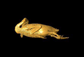
Is Radiology a Delicious Peking Duck?

not click on me


speaker.gif (1094 bytes)

ABSTRACT:
Preoperative evaluation in the living renal donor is essential. Recently 3-dimensional (D) MRI reconstructions with the advent of computer software provide enhanced visualization and can be interactive and more comprehensible as compared to 2D images and allow to provide consistent, accurate information concerning renal vascularity, especially venous anatomy, collecting system and parenchyma in preoperative evaluation of the living renal donor. Some of these reconstructions such as MIP have been used as the standard technique to visualize renal vascularity. Some of them can be color-coded and yo-yo moving displayed. Importantly the 3-dimensional (D) MRI reconstructions are excellent for the transplant surgeon and the clinic teaching. Currently several computer softwares have been provided to facilitate the 3D MRI reconstructions on a workstation.

speaker.gif (1094 bytes)

CLKHERE-1.gif (2771 bytes)